
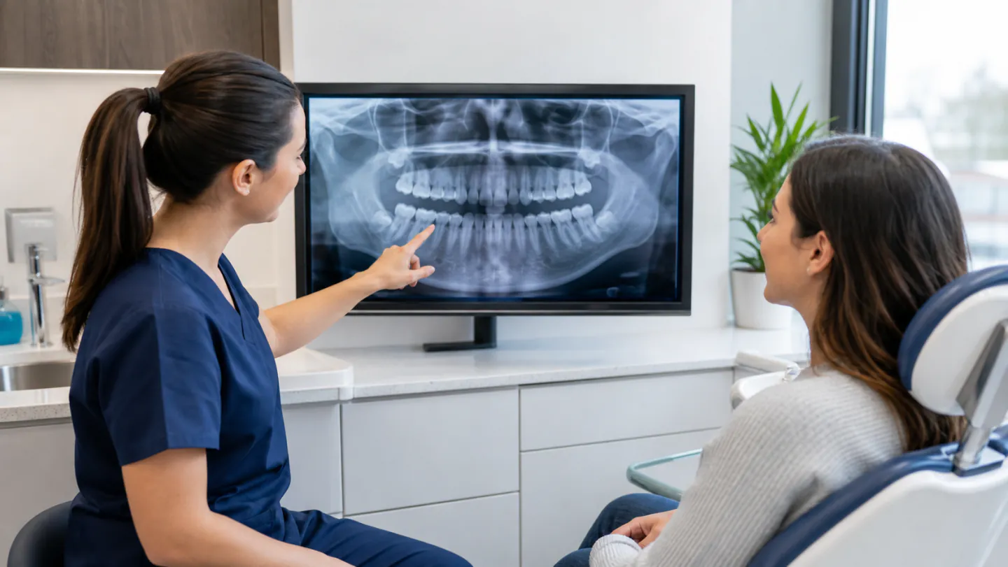
Dental X-rays can create mixed feelings. They help a dentist see what an examination cannot, but patients reasonably want to know why an image is needed and how much radiation is involved. “We always take these” is not a satisfying explanation.
An X-ray should answer a clinical question. It may show decay between teeth, bone levels around roots, infection, tooth development, or the position of a wisdom tooth. The type and timing should match your health, age, symptoms, previous images, and risk—not simply the date on the calendar.
If you have questions about dental X-rays in Winnipeg, this guide explains the common types, safety approach, and conversations to have before an image is taken.
Why the dentist cannot see everything directly
During an examination, the dentist can view tooth surfaces, gums, tongue, cheeks, and other visible tissues. They can feel for swelling, measure gum pockets, test teeth, and assess the bite.
They cannot directly see between tightly contacting teeth, inside a tooth, beneath the gum, or within the jawbone. Disease in those places may progress without a visible opening or pain.
An X-ray creates a two-dimensional picture based on how tissues absorb radiation. Dense material such as enamel and metal appears lighter. Less dense areas, including some decay and bone loss, appear darker. The image supports diagnosis but still needs interpretation alongside symptoms and examination findings.
Bitewing X-rays
Bitewings usually show the crowns of upper and lower back teeth and the bone height between them. The patient bites on a holder while the image is taken.
They are commonly used to look for cavities between teeth and under edges of fillings or crowns. They can also help monitor bone levels affected by gum disease.
The number and frequency depend on age, mouth size, cavity history, existing dental work, and risk. A person with several new cavities and dry mouth may need monitoring sooner than an adult who has remained stable for years.
Periapical X-rays
A periapical image shows a smaller group of teeth from crown to root tip and the surrounding bone. It is useful when the dentist is assessing pain, infection, trauma, root shape, or a particular tooth.
These images may be taken during root canal treatment, before some extractions, or to monitor an area near a root. A normal-looking crown does not rule out an infection at the tip, and a dark area does not diagnose a problem by itself.
The dentist compares the image with nerve tests, tapping, gum findings, symptoms, and older records when available.
Panoramic X-rays
A panoramic machine rotates around the head to create a wide view of both jaws, teeth, jaw joints, and nearby structures. The image is useful for general development, impacted teeth, large areas of the jaw, and some surgical or denture planning.
It does not show tiny cavities between teeth as clearly as bitewings. One large image is not a substitute for every smaller view.
During the scan, you bite on a guide and remain still while the machine moves. Positioning matters; jewellery, movement, or incorrect head angle can create distortion.
Three-dimensional cone-beam scans
A cone-beam computed tomography scan, often shortened to CBCT, creates a three-dimensional view of teeth, bone, and nearby structures. It may be used for implant planning, complicated root treatment, impacted teeth, surgical assessment, or other specific needs.
A CBCT provides more information and generally more radiation than a small two-dimensional dental X-ray. It should therefore be selected when the three-dimensional detail is expected to change diagnosis or treatment.
Ask which area will be scanned, why a regular image is not enough, and who will interpret the complete volume. More information is not automatically better when it does not affect care.
Digital X-rays and film
Most modern dental offices use digital sensors or plates. The image appears on a screen, can be enlarged, and can be transferred with the patient’s authorization. Digital systems avoid chemical film processing and can work with low exposure settings.
The word “digital” does not remove the need for careful technique. The correct sensor, exposure, positioning, and reason are still important.
Ask the dentist to show you the image. Seeing the area between teeth or around a root can make the treatment conversation easier to understand. The image belongs in the diagnostic record, not only on a monitor that the patient never sees.
How often are dental X-rays needed?
There is no universal schedule for everyone. The Canadian Dental Association explains that a healthy adult without recent cavities may not need X-rays at every appointment, while a less stable situation may require more frequent monitoring.
Frequency can be affected by:
- New or recurring cavities
- Dry mouth
- Gum disease and bone loss
- Age and tooth development
- Symptoms or trauma
- Existing crowns, bridges, implants, or large fillings
- Planned treatment
- Previous images and how recent they are
- Medical conditions that change oral risk
The useful question is, “What do you need to learn from this image today?”
Radiation and the benefit-risk decision
Radiation is part of the environment and many medical imaging procedures. Dental images use a small, focused amount, but exposure should still be justified and kept as low as reasonably achievable while producing a useful image.
Health Canada’s medical and dental X-ray guidance emphasizes that imaging should be necessary and appropriate and optimized to obtain the needed information with the lowest suitable dose.
The benefit is earlier or more accurate diagnosis. The risk of a properly selected image is small, but unnecessary repeat images provide no benefit. Good practice is not “never take X-rays” or “take a full set every year.” It is choose the right image for the right reason.
Why a dental X-ray might need to be repeated
Movement, a bent sensor, overlapping teeth, or a positioning error can hide the area the dentist needs to see. In that case, a repeat may be necessary. The decision should be based on missing diagnostic information, not on making every image look perfect.
Tell the team if sensors are painful or trigger gagging. Smaller holders, slower positioning, breathing through the nose, a more upright chair, or another image type may help.
If a repeat is suggested, it is reasonable to ask what was not visible. A respectful explanation takes only a moment.
Dental X-rays during pregnancy
Tell the clinic if you are pregnant or could be pregnant. Routine images that are not currently needed may be postponed. If you have pain, swelling, trauma, or suspected infection, the information from a necessary image can be important for safe treatment.
The dentist weighs urgency, area, and available alternatives and uses appropriate exposure controls. Do not delay an emergency assessment solely because you assume no image can be taken.
Ask what the image will change and whether an older image can help. Your dentist can also coordinate with your prenatal provider when medical circumstances are complex.
Dental X-rays for children
Children are not simply small adults. Their mouths are developing, and the type and timing of images depend on age, tooth contact, cavity risk, growth, and symptoms.
X-rays may help find cavities between baby teeth, monitor permanent tooth development, assess injury, or plan orthodontic care. A child with open spaces between teeth and low cavity risk may need different images from a child with tight contacts and several previous cavities.
Parents can ask why each image is recommended. Avoid frightening language about radiation in front of the child. Calmly explain that the picture helps the dentist see places hidden between and under teeth.
Do you need new X-rays when changing dentists?
Not always. Recent images can often be transferred with your permission if they are current, readable, and show what the new dentist needs.
Ask your previous clinic to send records before the first visit. The new dentist will review the quality, date, and your current health. Additional images may still be needed if symptoms have changed, an area was not captured, or the previous image is no longer diagnostic.
The CDA’s position on dental radiography encourages obtaining suitable recent images rather than automatically prescribing new ones only because a patient changed offices.
Can you refuse a dental X-ray?
You have the right to ask questions and decline. The dentist also has a responsibility to diagnose and treat safely. If an essential image is refused, they may be unable to provide a particular treatment or confirm that an area is healthy.
A useful conversation covers the reason, benefit, exposure, alternatives, and consequence of not taking it. “No X-rays ever” may leave serious problems hidden. “Whatever the office normally does” may expose you without enough individual thought. Informed choice sits between those extremes.
If you remain uncomfortable, ask whether the decision can be deferred, whether previous images are available, or whether a second opinion would help.
What X-rays cannot show perfectly
An image is not a photograph of disease. Early cracks may not appear. A cavity can look smaller on an X-ray than it is in the tooth. Two-dimensional views can overlap structures. Some dark and light areas are normal anatomy.
That is why dentists do not diagnose from an image alone. Symptoms, direct examination, history, testing, and changes over time all matter.
Be cautious with anyone—professional or online—who points to one shadow and promises a certain diagnosis without the rest of the clinical information.
X-ray costs and coverage in Winnipeg
Fees depend on the type and number of images. Small intra-oral images, panoramic views, and three-dimensional scans are different services.
Private insurance and the Canadian Dental Care Plan may contribute according to frequency limits, diagnostic rules, and fee schedules. A plan may decline an image because of timing even when the dentist believes it is clinically useful. Coverage and need are separate questions.
Ask for an estimate for larger scans and whether referral or interpretation fees are included. Bring previous records to reduce avoidable duplication.
Questions to ask before a dental X-ray
- What are you looking for?
- How will the result affect my treatment?
- Do you have a recent image that already shows this area?
- Why is this type of X-ray the best choice?
- Is the image urgent or can it wait?
- What can we do if I have a strong gag reflex?
- Are there special considerations for pregnancy or my health?
- May I see the image afterward?
Questions are part of informed care, not an inconvenience.
Dental X-rays in Winnipeg
The best dental imaging is selective. It reveals information that matters, uses appropriate exposure, and supports a diagnosis that is also based on you—not only a picture.
Winnipeg North Dental uses dental technology as a support for clinical judgment. If you have pain, are due for a check-up, or want to understand why an image has been recommended, request an appointment for an examination and a clear explanation.